6 December 2016
Scientists in Portugal have identified a potential biomarker that can gauge the severity of chronic heart failure using an advanced type of scanning probe microscope.
Cardiovascular diseases are the leading cause of mortality worldwide, responsible for about one-third of all deaths. Ischaemic cardiomyopathy is the most common cause of heart failure. It involves weakening of the heart muscle that can cause blood clots, cutting off the main blood supply to the heart.
High levels of fibrinogen, a protein common in the bloodstream and essential for clot formation, are a potential risk factor for cardiovascular diseases, but the underlying biological mechanisms remain unclear. Increased aggregation of erythrocytes, or red blood cells, has been linked to high levels of fibrinogen.
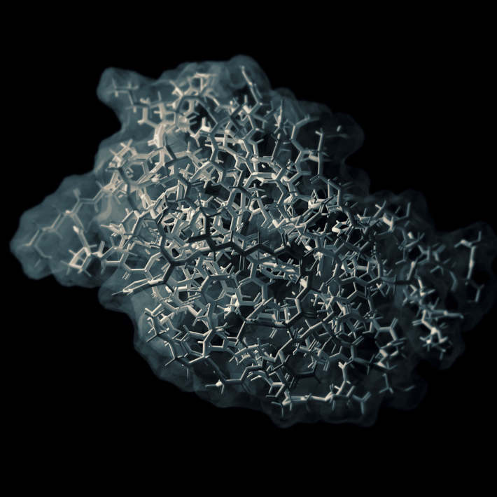
The team of scientists scrutinized the interaction between fibrinogen and erythrocytes. They used atomic force microscopy (AFM), a very high-resolution microscopy to the order of fractions of a nanometre, to examine how red blood cells aggregate in chronic heart failure patients.
“We show that the force required to break the binding between fibrinogen and erythrocytes is higher in patients with chronic heart failure than in healthy people,” says biochemist Nuno Santos from Universidade de Lisboa. The bonds were also stronger in patients with ischaemic chronic heart failure compared to non-ischaemic patients.
Chronic heart failure patients with higher fibrinogen-erythrocyte binding forces were hospitalized more frequently in the 12 months that followed the initial assessments.
“This technique has also allowed us to study red cell stiffness. Erythrocytes from these patients showed changes in their elasticity and behaviour while in the blood stream,” says Santos. These changes are thought to have an effect on the strength of the bond red cells form with fibrinogen.
Santos praises the potential of AFM in disease severity identification, labelling it “an important advancement in the field of nanotechnology for cardiology.”
He acknowledges, however, that the study is limited by the relatively small number of cohorts.
The study shows how nano-based technologies can be used to identify cardiovascular problems in a field where biomarkers relevant to cardiovascular risk still have limited applicability.
References
- Guedes, A. F., Carvalho, F. A., Malho, I., Lousada, N., Sargento, L., et al. Atomic force microscopy as a tool to evaluate the risk of cardiovascular diseases in patients. Nature Nanotechnology. (2016). | article