25 December 2016
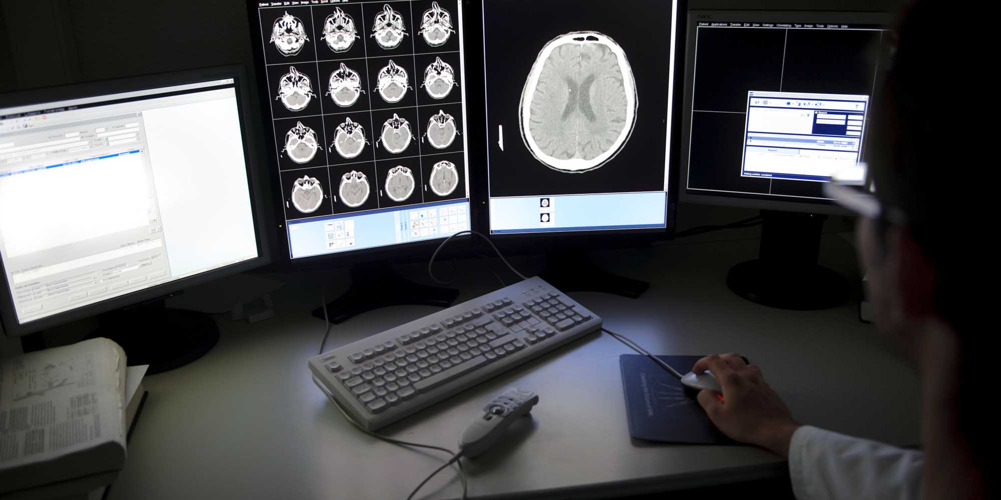
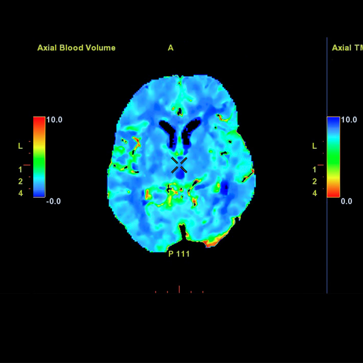
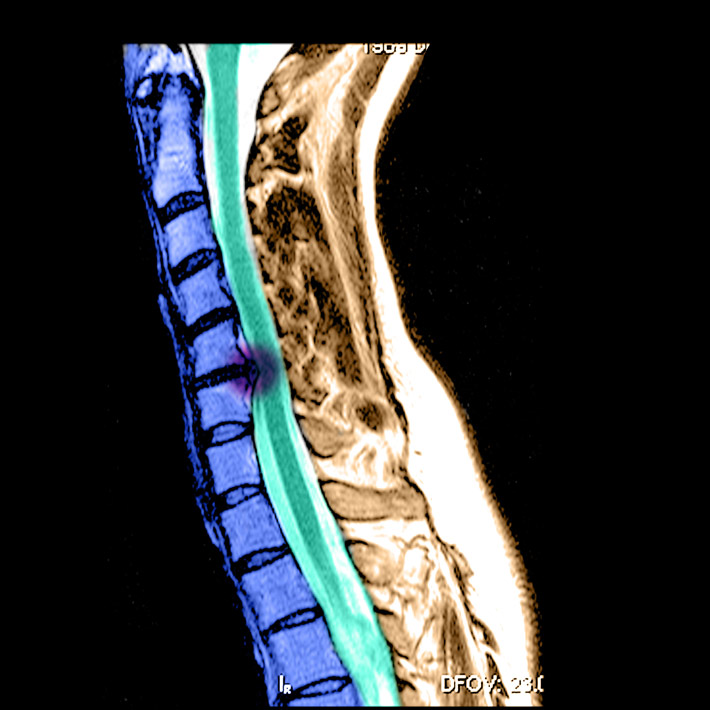
New sophisticated imaging algorithms have allowed clinicians to reduce patient exposure to radiation while maintaining the quality of the images from CT scans. But researchers warn these algorithms have their limits, compromising image quality and diagnosis.
Since 2011, clinicians at Saudi Arabia’s Ministry of National Guard—Health Affairs (MNGHA) have been using an image reconstruction algorithm called adaptive statistical iterative reconstruction (ASIR) to reduce image ‘noise’ in CT scans and improve image quality.
Image noise can be thought of as the visual equivalent of the subtle background hiss in old radios. On a CT image, noise appears as random speckles that can significantly reduce image quality, making it more difficult to pick up lesions or abnormalities on a scan.
Achieving a sharp image has always been a challenge for radiologists. Traditionally, they used a method called filter back projection (FBP) to reduce image noise.
Advances in computer technologies have enabled the use of complex mathematical models, like ASIR, to provide good image quality at reduced radiation doses. This is particularly important for people needing to undergo a series of CT scans. These models learn from the data they receive from the X-ray machine and reconstruct those parts of the CT images affected by noise.
The Saudi researchers noticed reduced sharpness on some ASIR images, so they decided to assess the impact of ASIR on image quality to make sure the technique was not compromising the diagnostic information on the CT images. They measured image quality obtained at various radiation doses using both conventional FBP and ASIR.
They discovered that while ASIR undoubtedly offered an improved image quality, this was dependent on the level of radiation exposure applied by the radiologist and the percentage level of ASIR applied in the scan.
In other words, ASIR improved the resolution of a scan up to a certain threshold, beyond which it started to degrade the quality of the image.
“This threshold changes to higher or lower values depending on the scanning dose used to obtain the image,” write the researchers in their study published in the Journal of Applied Clinical Medical Physics.
When the resolution or sharpness of an image starts degrading, some information that may be clinically significant is removed from the image, they add. “Physicians need to be careful when acquiring CT images using ASIR”.
As a result, the team now avoids the use of higher levels of ASIR.
More research is needed, they say, to identify the exact anatomical information that is lost when the sharpness of the images starts to degrade.
“I believe [the findings] will make scientists in the field re-think when and how to use ASIR and similar algorithms,” says Fahad Ahmed Hussain of MNGHA.
References
Hussain, F., Mail, N., Shamy, A., Alghamdi, S. & Saoudi, A. A qualitative and quantitative analysis of radiation dose and image quality of computed tomography images using adaptive statistical iterative reconstruction. Journal of Applied Clinical Medical Physics. 17, 419-432 (2016).| article


