6 September 2017
Most serious diseases present three key challenges: diagnosis, targeted treatment, and monitoring of disease progression and response to therapy. KAIMRC researchers, led by nanoscientist Khalid Abu-Salah, with colleagues in Sweden and the US have developed nanoparticles that might perform these roles simultaneously1.
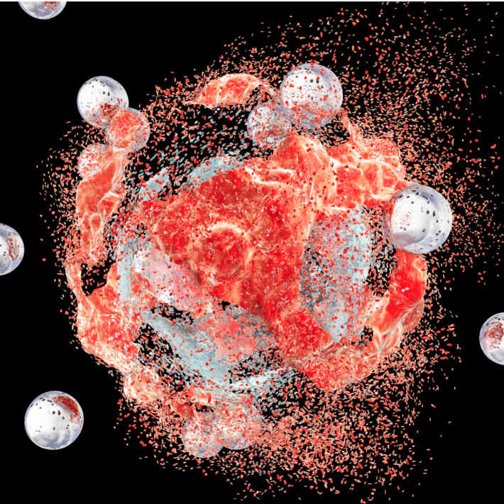
Their system is based on therapeutic and diagnostic nanocarriers built of tiny spherical structures called micelles, composed of poly-ethyleneglycol (PEG) and poly-caprolactone (PCL) polymers. The micelles have been used as vehicles to carry the anti-cancer drug busulphan along with iron oxide particles that make them visible in MRI scans. They can also be labelled with fluorescent dyes to become readily detectable under suitable illumination.
They are compatible with biological tissues, promoting no adverse reactions such as inflammation, and have biodegradable properties that assist their excretion after they have fulfilled their mission. The researchers hope to be able to target specific sites of disease by coating the micelles with molecules, such as antibodies, that selectively bind to receptors on the surface of diseased cells.
The combined drug delivery and imaging system has been tested in mice. These animal trials were complemented by tests with cultured cells that demonstrated efficient release of the drug over timescales that could prove clinically useful.
Abu-Salah is keen to target breast cancer, which is affecting more women in Saudi Arabia every year. “We will investigate the potential on animals with breast cancer and leukaemia, then hopefully move onto tests with cancer patients,” he says.
“The multi-functional ability is the key advantage of our system.”
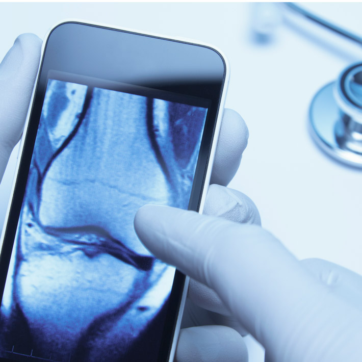
The ability to follow the distribution of the micelles by MRI and fluorescence imaging techniques will be used to find the most efficient method to get the micelles to selectively target diseased tissues. Such targeting could also prove useful to indicate if a cancer, for example, is shrinking after treatment and to reveal the extent to which a disease has spread to other regions of the body. Once any targeting procedure has been validated it could be used in the early stages of clinical investigation to diagnose where a specific cancer is located and how far it has advanced.
“We hope our work will eventually help to treat cancer with minimal side effects on healthy cells,” says Abu-Salah.
Their system is based on therapeutic and diagnostic nanocarriers built of tiny spherical structures called micelles, composed of poly-ethyleneglycol (PEG) and poly-caprolactone (PCL) polymers. The micelles have been used as vehicles to carry the anti-cancer drug busulphan along with iron oxide particles that make them visible in MRI scans. They can also be labelled with fluorescent dyes to become readily detectable under suitable illumination.
They are compatible with biological tissues, promoting no adverse reactions such as inflammation, and have biodegradable properties that assist their excretion after they have fulfilled their mission. The researchers hope to be able to target specific sites of disease by coating the micelles with molecules, such as antibodies, that selectively bind to receptors on the surface of diseased cells.
The combined drug delivery and imaging system have been tested in mice. These animal trials were complemented by tests with cultured cells that demonstrated efficient release of the drug over timescales that could prove clinically useful.
Abu-Salah is keen to target breast cancer, which is affecting more women in Saudi Arabia every year2. “We will investigate the potential on animals with breast cancer and leukaemia, then hopefully move onto tests with cancer patients,” he says.
“The multi-functional ability is the key advantage of our system.”
The ability to follow the distribution of the micelles by MRI and fluorescence imaging techniques will be used to find the most efficient method to get the micelles to selectively target diseased tissues. Such targeting could also prove useful to indicate if a cancer, for example, is shrinking after treatment and to reveal the extent to which a disease has spread to other regions of the body. Once any targeting procedure has been validated it could be used in the early stages of clinical investigation to diagnose where a specific cancer is located and how far it has advanced.
“We hope our work will eventually help to treat cancer with minimal side effects on healthy cells,” says Abu-Salah.
References
- Asem, H., Zhao, Y., Ye, F., Barrefelt, Å., Manuchehr, A-V. et al. Biodistribution of biodegradable polymeric nano-carriers loaded with busulphan and designed for multimodal imaging. Journal of Nanobiotechnology 14, 82 (2016). | article
- Saggu, S., Rehman, H., Abbas, Z. K. & Ansari, A. A. Recent incidence and descriptive epidemiological survey of breast cancer in Saudi Arabia. Saudi Medical Journal 36, 1176-80 (2015).| article