5 April 2020
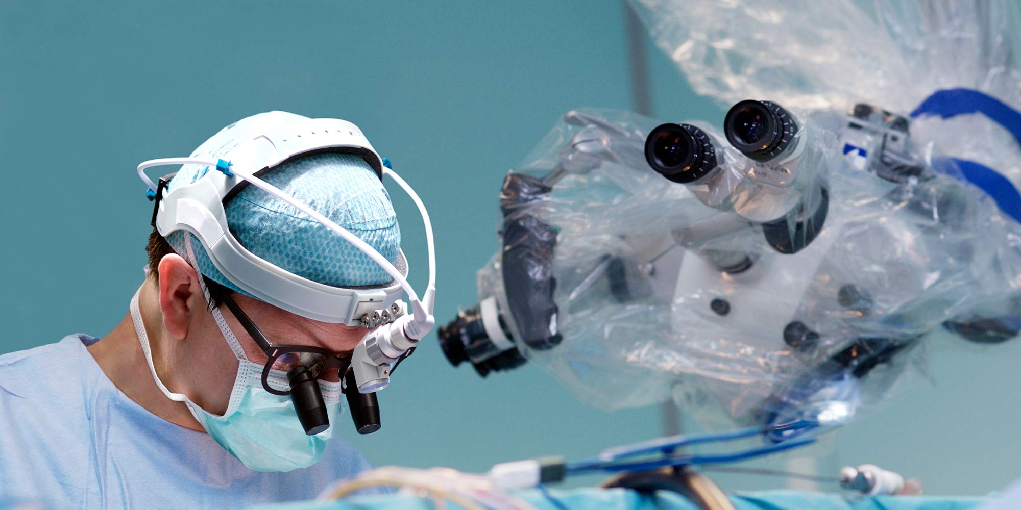
Artificial intelligence can provide surgeons with near real-time diagnostic information during removal of brain tumors, a study published in Nature Medicine has shown. The work, led by Daniel Orringer, a neurosurgeon at NYU Langone Health, USA, could improve surgical care of brain tumours.
During surgery to remove a brain tumour, analysis of resected tissue is essential to identify the type of tumour and to ensure that as much cancerous tissue as possible is removed while minimizing removal of healthy tissue. The conventional process involves transferring samples to a laboratory for staining and analysis by a pathologist, but this is time-consuming, expensive and limited by resources and expertise.
Technological advances have led to the introduction of stimulated Raman histology (SRH), a technique that enables simple, rapid microscopic analysis of tissue samples in the operating room. “SRH offers the surgeon the ability to see a tumour at a microscopic scale that would otherwise be invisible and to rapidly diagnose the type of tumour they’re operating on,” explains Orringer. “However, while SRH rapidly produces high-resolution microscopic images, the interpretation of those images still requires considerable expertise.”
Orringer and colleagues aimed to overcome this limitation by developing a system for automated analysis of SRH images. They used artificial intelligence to generate deep convolutional neural networks that could analyze samples. These networks were trained with more than 2.5 million classified images of tumour samples to identify tumour subtypes on the basis of distinguishing characteristics.
The team tested their system with a series of 278 patients who underwent surgery for a brain tumour. Samples from each patient were analyzed with both the automated system and by a pathologist, and the accuracy of the techniques was compared. With the automated system, 94.6% of tumours were accurately classified, compared with 93.9% with the conventional technique. The automated result was also generated approximately ten times faster, typically within 2.5 minutes. “Our system could reliably predict diagnosis in brain tumour patients in minutes with accuracy comparable to that of expert neuropathologists,” summarizes Orringer.
The system has clear implications for improving diagnosis and tumour removal, effectively offering immediate diagnostic information to surgeons in the operating room. “To some extent, it’s like having the expertise of a pathologist there, even if the pathologist cannot physically be present,” explains Orringer. “This is particularly important in settings where the expertise of a pathologist is needed but a lab, a pathologist or infrastructure, are lacking.”
References
- Hollon, T.C., Pandian, B., Adapa, A.R. et al. Near real-time intraoperative brain tumor diagnosis using stimulated Raman histology and deep neural networks. Nat Med 26, 52–58 (2020). | article