14 August 2016
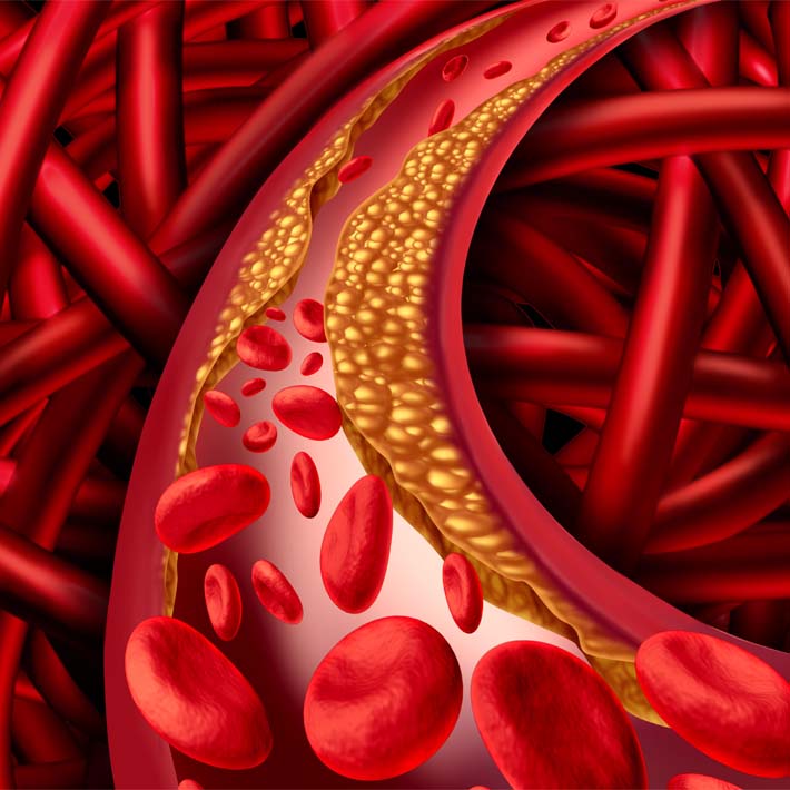
Atrophy is a common consequence of both congestive heart failure and cancer, as skeletal muscle throughout the body undergoes a process of steady degeneration. Researchers have now identified a cellular mechanism associated with this deterioration, which could offer a useful drug target for preventing muscle wasting1.
Angiotensin II has been identified by scientists, led by Jens Fielitz at Charité Universitätsmedizin Berlin in Germany, as a specific trigger for atrophy in congestive heart failure patients. In the aftermath of cardiac damage, large quantities of this hormone are released into the bloodstream. As it circulates, it sets molecular events in motion that result in selective destruction of muscle proteins. A protein called muscle RING-finger-1 (MuRF1) is a critical link in this chain, serving as the enzyme that directly marks doomed proteins for elimination.
In an effort to identify the intermediate steps in this process, Fielitz and colleagues conducted a search for proteins that can switch on the gene encoding MuRF1. This led them to a protein known as transcription factor EB (TFEB), which is produced in high levels in both skeletal and heart muscle, and binds to DNA sequences in the genome that are responsible for regulating MuRF1 production. Intriguingly, TFEB has also been connected with the control of other well-known protein degradation pathways in previous research, although this study represents the first direct link between this molecule and muscle atrophy.
Subsequent experiments further strengthened this association. Under normal conditions, muscle cells have inhibitory processes in place that prevent TFEB from stimulating the production of MuRF1. However, when angiotensin II is present in the bloodstream, it triggers specific cellular events that alleviate this inhibition. As a result, MuRF1 levels rise and the process of atrophy begins. Accordingly, the researchers showed that they could thwart angiotensin II-mediated atrophy in cultured muscle cells by directly reducing TFEB levels or selectively eliminating the molecules that relieve the inhibition of TFEB.
The authors note that muscular atrophy is the product of many complex physiological processes, including prolonged physical inactivity in congestive heart failure patients. Nevertheless, the central role of angiotensin II is well established, and the authors see strong evidence from their experiments that “TFEB played a key role in regulating angiotensin II-induced skeletal muscle atrophy.”
If these findings can be further validated in animal models, the regulatory processes that control TFEB activity might ultimately offer a worthy target for drugs aimed at slowing or reversing the muscle-wasting process.
References
- Cheng, W., Pullin, D. I., & Samtaney, R. Angiotensin II Induces Skeletal Muscle Atrophy by Activating TFEB-Mediated MuRF1 Expression. Circ. Res. (2015). | article