6 December 2016
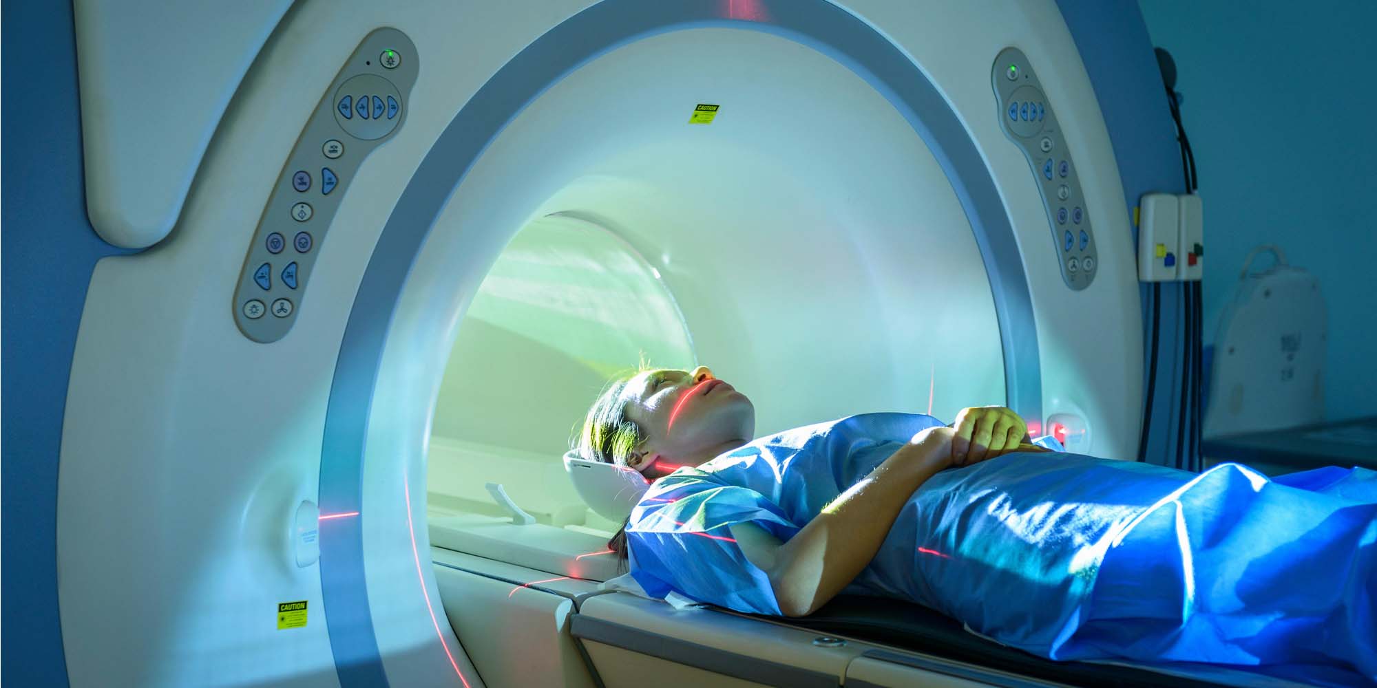
Magnetic resonance imaging (MRI) enables physicians to see inside bodies in great detail without invasive surgery. Contrast agents can further extend the capacity of MRI to include cancer diagnosis by reacting to changes associated with lesions and tumours, such as an increase in the surrounding acidity.
A pH figure is used to express a solution’s acidity or alkalinity. Figures above seven signify an alkaline solution, while those below seven signify an acidic solution.
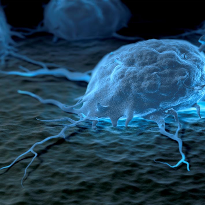
Kazunori Kataoka at the University of Tokyo and co-workers1 have now developed a contrast agent that amplifies magnetic resonance signals when the surrounding pH drops below 6.7. If injected into patients, the contrast agent could illuminate tumours and improve the contrast of their pathological features on MRI scans.
The researchers prepared pH-sensitive nanoparticles — the crucial ingredient in this newly developed contrast agent — by encapsulating manganese ions and calcium phosphate fragments in polyethylene glycol shells. The manganese ions are designed to amplify magnetic resonance imaging, while the calcium phosphate fragments are designed to bind manganese ions under normal conditions and subsequently release them when the pH drops below a critical value.
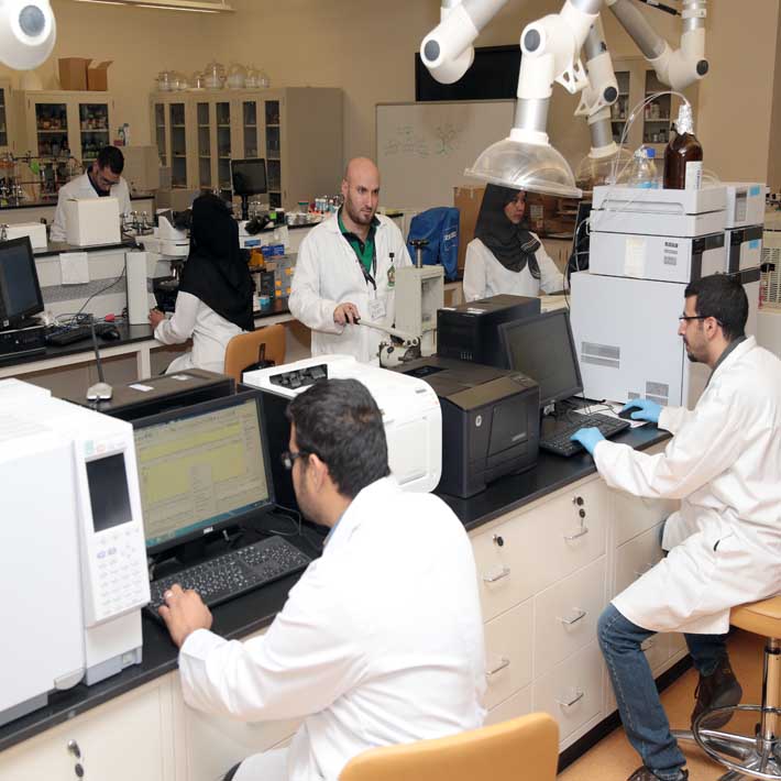
Kataoka and his team examined how these nanoparticles behave at different pH ranges, including pH 5–6 (intracellular conditions), pH 6.5–6.8 (tumour microenvironment) and pH 7.4 (blood). They found that after four hours, 95%, 71% and 36% of manganese ions were discharged from the nanoparticles at pH 5.0, 6.5 and 6.7 respectively. In comparison, less than 8% of manganese ions were released at pH 7.4.
The researchers then tested the contrast capability of their nanoparticles in tumour-bearing mice. They found that the tumour/normal tissue (T/N) ratio of the nanoparticles reached 130% within 30 minutes after administration through intravenous injection. The high T/N ratio lasted for hours, providing sufficient time for MRI scans to be taken. Altogether, the results show that the nanoparticles rapidly and selectively bind solid tumours and brighten them up through contrast enhancement.
Lastly, Kataoka and his team checked whether their nanoparticles could trace spreading tumours. They applied the nanoparticles to a mouse liver that had been tainted with colon cancer cells. The spreading tumours lit up and revealed their migratory paths within one hour of administration through intravenous injection. The team reports that its nanoparticles were thus useful both for cancer diagnosis and the visualisation of metastases.
References
Mi, P., Kokuryo, D., Cabral, H., Wu, H., Terada, Y. et al. A pH-activatable nanoparticle with signal amplification capabilities for non-invasive imaging of tumour malignancy. Nature Nanotechnology (2016). |article