11 August 2016
Magnetic metal oxide nanoparticles have demonstrated considerable promise for a wide variety of biomedical applications, but researchers have struggled to identify an optimal strategy for producing them.
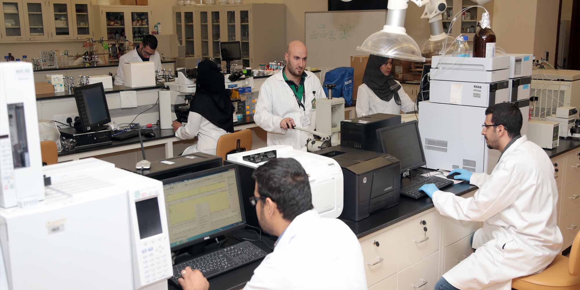
Researchers, led by nanobiochemist Kheireddine El-Boubbou of King Abdullah International Medical Research Center, have now developed an approach for producing these particles easily, cheaply and safely.
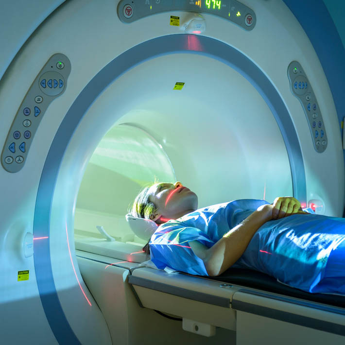
“Tailored nanoprobes of controlled sizes and compositions based on iron oxides can be used for applications such as detection, imaging and drug delivery, both in vitro [in the body] and in vivo [in the lab],” says El-Boubbou. Unfortunately, most existing methods yield aggregation-prone nanoparticles that vary in size and shape, or require the use of costly and toxic chemicals. “The lack of simple, fast, large-scale and cost-effective preparation remains a major hurdle toward real-world biomedical applications,” he says.
El-Boubbou and his colleagues devised a strategy they call Ko-precipitation hydrolytic basic (KHB). It involves combining metal-based salts dissolved in water with different fatty acids in the presence of chemicals known as alkylamines. This forms highly stable acid-coated metal oxide nanocrystals. All of the materials involved are inexpensive and readily available, and the process can be performed at much lower temperatures (80 °C) than existing techniques, which require temperatures in the range of 200 to 300 °C.
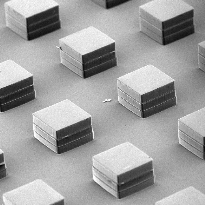
Electron microscopy confirmed that that the KHB method can readily produce highly homogeneous populations of iron-oxide nanoparticles. El-Boubbou and colleagues subsequently demonstrated that by modulating the ratio of fatty acid to iron salt solution, they could accurately control the size of the resulting clusters, consistently generating spherical nanoparticles with diameters ranging from two to ten nanometres.
This approach proved to extend to metals other than iron—including copper, nickel and zinc—widening the potential range of applications. The researchers were also able to modulate the chemical properties of the final product. The first batch of nanoparticles produced was not soluble in water, but by modifying their protocol slightly, they were able to generate iron oxide nanoparticles that are. These could prove more useful for applications that require the particles to function within the interior of living cells.
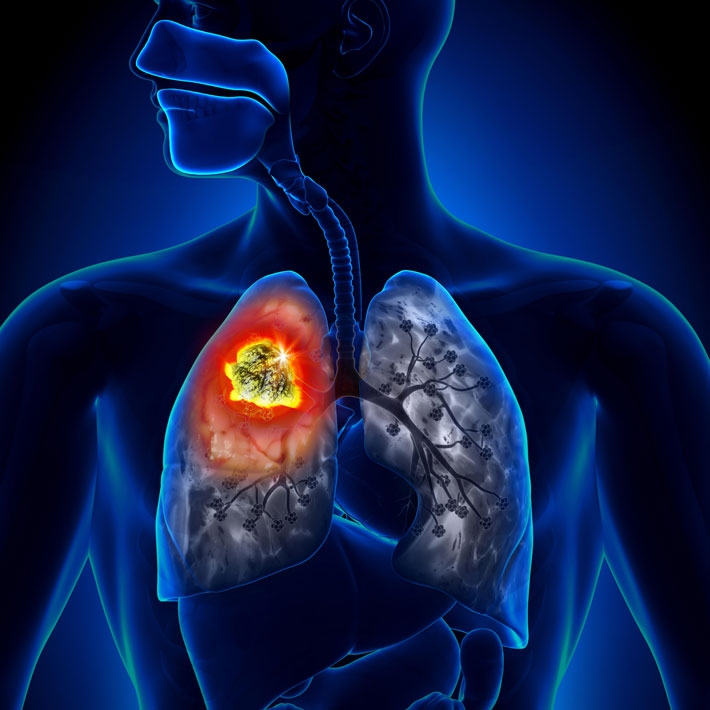
El-Boubbou has spent much of the past decade developing magnetic nanoprobes for biomedical applications. He has already begun to test the performance of these KHB-derived nanoparticles as a clinical research tool. “We will utilize these functional nanoparticles for cancer imaging and as carriers to deliver drugs to tumours in an attempt to develop chemotherapeutic approaches,” he says.
References
El-Boubbou, K., Al-Kaysi, R.O., Al-Muhanna, M.K., Bahhari, H.M., Al-Romaeh, A.I. et al. Ultra-small fatty acid-stabilized magnetite nanocolloids synthesized by in situ hydrolytic precipitation. Journal of Nanomaterials (2015). | article