10 March 2019
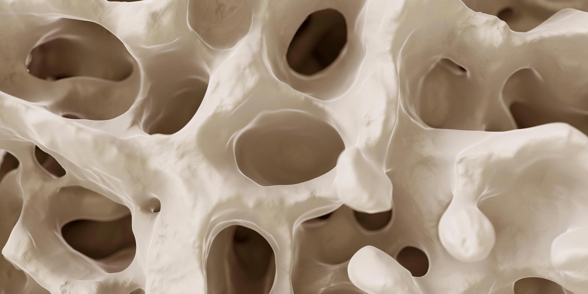
A team of scientists in Switzerland have mapped a comprehensive atlas of the cells and structures that make up the microenvironment of bone marrow.
Until now, conventional imaging methods, such as electron microscopy, have offered limited, two-dimensional glimpses of bone marrow cells. Due to the difficulty of keeping the tissue structure intact while preparing thin sections of bone for microscopic analysis, the spatial distribution of cells within the bone marrow has been unclear.
To obtain a better view, Timm Schroeder, and colleagues at the Department of Biosystems Science and Engineering, ETH Zürich, developed a 3D multicolour mapping method that has enabled them to visualise the bone marrow in whole leg bones (femurs) of adult mice.
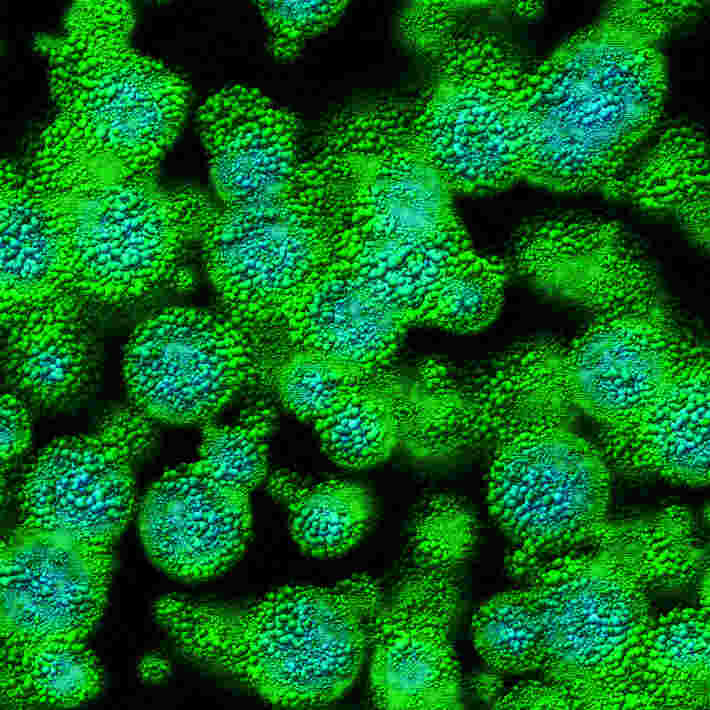
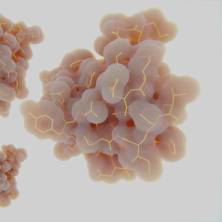
The researchers first used a technique called immunostaining, which involves using antibodies that can identify specific markers in biological tissue. They were then able to selectively visualise those markers via fluorescent labelling. From an initial list of nearly 250 antibodies, 67 were extensively tested by the team to choose the ones that best selectively stained the cell types they were analysing. The researchers then used an imaging method called multicolour quantitative confocal imaging cytometry to generate an atlas of more than 40 molecules, including markers, which indicate the precise distribution of particular cells of interest.
Specifically, they achieved state-of-the-art imaging of osteoblastic cells (which produce bone), vascular and perivascular cells (related to or surrounding blood vessels), neuronal cells (linked to the nervous system), and stromal cells (found in connective tissue). They also detected several types of extracellular matrix proteins, which are thought to provide structural support to surrounding cells.
Reporting their findings in Nature Biotechnology they state: “Although our results mainly confirm the conclusions of previous studies, they also extend some previous observations and contradict others.”
For example, a marker called Nes-GFP, which is known to indicate the presence of specialised nerve cells known as neuronal progenitors in the central nervous system, unexpectedly showed that those cells are also found in nerve fibres inside bone marrow. This is thought to be the first time that those cells have been identified outside the brain and spinal cord.
The study also revealed that vascular cells, perivascular cells and stromal cells may be more diverse than previously thought. For example, a stromal cell marker called CD271 indicated stromal cell activity throughout the bone marrow, with the highest amount present in its central part. Using a stain called boron dipyrromethene, the researchers found that bone marrow adipocytes, a type of stromal cell needed for metabolism, were mainly found in the distal parts of the bone marrow, although their number and location vary with age.
The researchers conclude that their observations “probably only scratch the surface of what can be gained from the data.” To enable the wider scientific community to delve deeper, they have made their imaging data freely available for download.
References
Coutu, D.L., Kokkaliaris, K.D., Kunz, L. & Schroeder, T. Three-dimensional map of nonhematopoietic bone and bone-marrow cells and molecules. Nature Biotechnology (2017). | article