26 August 2019
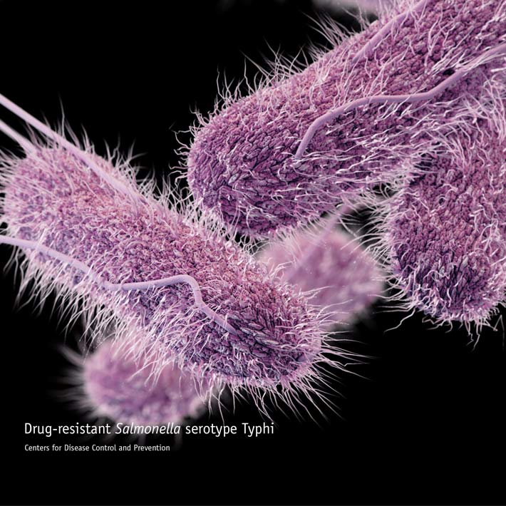
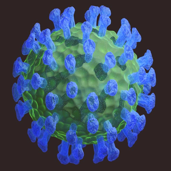
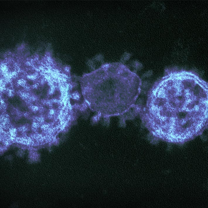
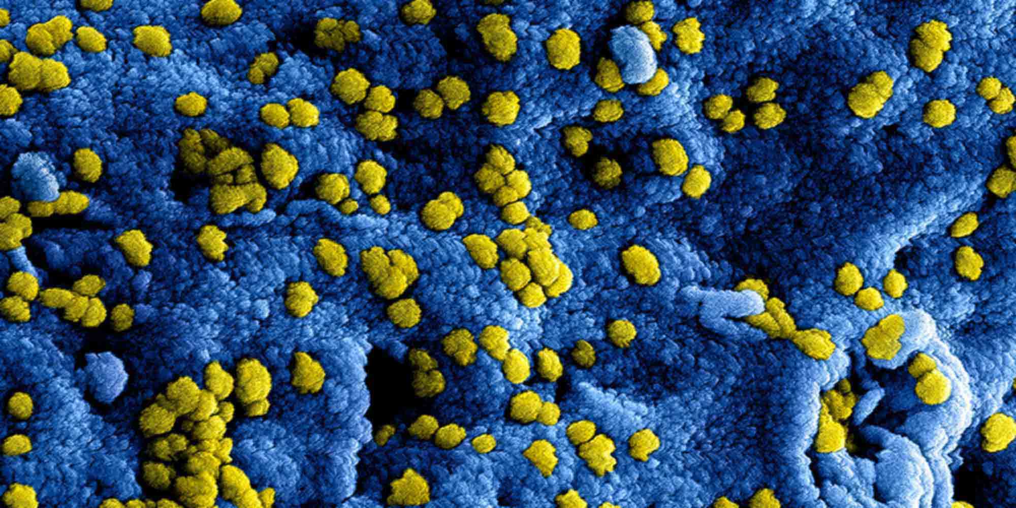
Patients suffering from infection with Middle East respiratory syndrome coronavirus (MERS-CoV) typically display a wide array of symptoms, ranging from a self-limiting cold-like illness to complete respiratory failure and death. However, little is known about the spread of the disease throughout the body, or which organs are predominantly affected.
Now, KAIMRC intensive care consultant Yaseen Arabi and co-workers have conducted an analysis of disease-related changes to key organs and of the dissemination of MERS-CoV viral particles in the body of a recently deceased patient. Their findings indicate that viral particles collect in various organs in the body, including the lungs and kidneys.
Organ dysfunction is a common element of severe MERS-CoV infection, particularly acute renal (kidney) failure, which is reported in 75 percent of critically ill patients. Alsaad’s team analysed organ tissue samples from a 33-year-old male patient who died in hospital following MERS-CoV infection in 2015. They collected samples from his heart, liver, kidneys, thigh muscles, brain and lungs within 45 minutes of death and conducted investigations using immunohistochemical staining and electron microscopy.
The lung tissues showed severe haemorrhagic pneumonia. The researchers found varying degrees of damage across the lungs, with worst affected areas containing large amounts of blood and fibrous mesh growths that impeded blood flow, together with disrupted air sac pores. The kidney tissue showed significant changes to tubular epithelial cells, which play a key role in kidney function. Some inflammation was found in the liver, but the team found no obvious changes to the heart and brain.
Alsaad’s team then traced accumulations of viral particles throughout the samples. They demonstrated that MERS-CoV is expressed in several cell types, including pneumocytes (cells responsible for gas exchange in the air sacs of the lungs), renal cells and the cells that line the interior of blood vessels. Renal cells appear to encourage MERS-CoV replication and generate significantly more infectious viral particles than bronchial cells. The researchers also identified viral particles entering skeletal muscle tissues via host macrophages.
Their findings suggest the possibility that the cells and tissues of the kidneys and skeletal muscles support the growth of the MERS-CoV virus, a phenomenon known as tropism.
“It is possible that our patient’s reaction to MERS-CoV infection was atypical [he also suffered from lymphoma],” say the team in their paper published in the journal Histopathology. “Further detailed studies are needed to explore the morphological changes associated with MERS-CoV infection, and ultrastructural as well as immunohistochemical localisation studies are required to further enhance our understanding of viral pathogenesis.”
References
Alsaad, K. O., Hajeer, A. H., Al Balwi, M., Al Moaiqel, M., Al Oudah, N. et al. Histopathology of Middle East respiratory syndrome coronavirus (MERS-CoV) infection – clinicopathological and unilateral study. Histopathology 72, 516–524 (2018). | article